Friday, November 16, 2018

Neurotransmitters

Definition

Neurotransmitters are chemicals located and released in the brain to allow an impulse from one nerve cell to pass to another nerve cell.

Neurotransmitters 772

Description

There are approximately 50 neurotransmitters identified. There are billions of nerve cells located in the brain, which do not directly touch each other. Nerve cells communicate messages by secreting neurotransmitters. Neurotransmitters can excite or inhibit neurons (nerve cells). Some common neurotransmitters are acetylcholine, norepinephrine, dopamine, serotonin and gamma aminobutyric acid (GABA). Acetylcholine and norepinephrine are excitatory neurotransmitters while dopamine, serotonin, and GABA are inhibitory. Each neurotransmitter can directly or indirectly influence neurons in a specific portion of the brain, thereby affecting behavior.

Mechanism of impulse transmission

A nerve impulse travels through a nerve in a long, slender cellular structure called an axon, and it eventually reaches a structure called the presynaptic membrane, which contains neurotransmitters to be released in a free space called the synaptic cleft. Freely flowing neurotransmitter molecules are picked up by receptors (structures that appear on cellular surfaces that pick up molecules that fit into them like a "lock and key") located
in a structure called the postsynaptic membrane of another nearby neuron. Once the neurotransmitter is picked up by receptors in the postsynaptic membrane, the molecule is internalized in the neuron and the impulse continues. This process of nerve cell communication is extremely rapid

Once the neurotransmitter is released from the neurotransmitter vesicles of the presynaptic membrane, the normal movement of molecules should be directed to receptor sites located on the postsynaptic membrane. However, in certain disease states, the flow of the neurotransmitter is defective. For example, in depression, the flow of the inhibitory neurotransmitter serotonin is defective, and molecules flow back to their originating site (the presynaptic membrane) instead of to receptors on the postsynaptic membrane that will transmit the impulse to a nearby neuron.
The mechanism of action and localization of neurotransmitters in the brain has provided valuable information concerning the cause of many mental disorders, including clinical depression and chemical dependency, and in researching medications that allow normal flow and movement of neurotransmitter molecules.

Neurotransmitters are chemicals that transmit messages from one nerve cell (neuron) to another. The nerve impulse travels from the first nerve cell through the axon—a single smooth body arising from the nerve cell— to the axon terminal and the synaptic knobs. Each synaptic knob communicates with a dendrite or cell body of another neuron, and the synaptic knobs contain neurovesicles that store and release neurotransmitters. The synapse lies between the synaptic knob and the next cell. For the impulse to continue traveling across the synapse to reach the next cell, the synaptic knobs release the neurotransmitter into that space, and the next nerve cell is stimulated to pick up the impulse and continue it.


Neurotransmitters are chemicals that transmit messages from one nerve cell (neuron) to another. The nerve impulse travels from the first nerve cell through the axon—a single smooth body arising from the nerve cell— to the axon terminal and the synaptic knobs. Each synaptic knob communicates with a dendrite or cell body of another neuron, and the synaptic knobs contain neurovesicles that store and release neurotransmitters. The synapse lies between the synaptic knob and the next cell. For the impulse to continue traveling across the synapse to reach the next cell, the synaptic knobs release the neurotransmitter into that space, and the next nerve cell is stimulated to pick up the impulse and continue it.

Neurotransmitters, mental disorders, and medications

Schizophrenia

Impairment of dopamine-containing neurons in the brain is implicated in schizophrenia , a mental disease marked by disturbances in thinking and emotional reactions. Medications that block dopamine receptors in the brain, such as chlorpromazine and clozapine , have been used to alleviate the symptoms and help patients return to a normal social setting.

Depression

In depression, which afflicts about 3.5% of the population, there appears to be abnormal excess or inhibition of signals that control mood, thoughts, pain, and other sensations. Depression is treated with antidepressants that affect norepinephrine and serotonin in the brain. The antidepressants help correct the abnormal neurotransmitter activity. A newer drug, fluoxetine (Prozac), is a selective serotonin reuptake inhibitor (SSRI) that appears to establish the level of serotonin required to function at a normal level. As the name implies, the drug inhibits the re-uptake of serotonin neurotransmitter from synaptic gaps, thus increasing neurotransmitter action. In the brain, then, the increased serotonin activity alleviates depressive symptoms.

Alzheimer's disease

Alzheimer's disease , which affects an estimated four million Americans, is characterized by memory loss and the eventual inability for self-care. The disease seems to be caused by a loss of cells that secrete acetylcholine in the basal forebrain (region of brain that is the control center for sensory and associative information processing and motor activities). Some medications to alleviate the symptoms have been developed, but presently there is no known treatment for the disease.

Generalized anxiety disorder

People with generalized anxiety disorder (GAD) experience excessive worry that causes problems at work and in the maintenance of daily responsibilities. Evidence suggests that GAD involves several neurotransmitter systems in the brain, including norepinephrine and serotonin.

Attention-deficit/hyperactivity disorder

People affected by attention-deficit/hyperactivity disorder (ADHD) experience difficulties in the areas of attention, overactivity, impulse control, and distractibility. Research shows that dopamine and norepinephrine imbalances are strongly implicated in causing ADHD.

Others

Substantial research evidence also suggests a correlation of neurotransmitter imbalance with disorders such as borderline personality disorders schizotypal personality disorder avoidant personality disorder social phobia histrionic personality disorder , and somatisation disorder .


Wednesday, November 14, 2018

ECG / EKG Components

Components of ECG


Each ECG cycles consists of 5 waves: P, Q, R, S, T corresponding to different phases of the heart activities. The P wave represents the normal atrium (upper heart chambers) depolarization; the QRS complex (one single heart beat) corresponds to the depolarization of the right and left ventricles (lower heart chambers); the T wave represents the re-polarization (or recovery) of the ventricles. To interpret ECG, one needs to focus on the frequency (heart rate), regularity, shape and size of each individual waves and the timing and interaction between waves. The following diagram shows the components of a ECG cycle.


P Wave

The P wave occurs when both left and right atria are full of blood and the SA node fires. The signal causes both atria to contract and pump blood to the ventricles (lower chambers). Any abnormality is generally associated with the SA node and the atria.
Lead II and V1 are the best leads to observe p-waves.
General characteristics :
  • Less than 100ms in duration
  • Less than 2.5mm in amplitude
Examples of abnormal P waves
Abnormal PatternsPossible Causes
Inverted
  1. Atrial depolarization is in a different direction
  2. Sinoatrial block: Pacemaker is no longer the SA node and is subsumed by another part of the heart.
  3. The heart is in an abnormal location or orientation within the chest (dextrocardia).
Greaten than 2.5mm in amplitude
  1. Right atrial enlargement
Longer than 100ms with two lobes
  1. Left atrial enlargement
Invisible or absent
  1. Impairment of conduction at the sinoatrial node.
  2. Sinoatrial block: SA node fails to discharge and AV junction takes over as the pacemaker.

PQ Segment

When the signal from the SA node arrives the AV node, the signal is slowed and paused for a short period to allow blood from the atria to fill the ventricles.
General characteristics :
  • 120 - 200ms in duration from the beginning of P wave to the beginning of QRS complex (PR interval)
Examples of abnormal PR interval
Abnormal PatternsPossible Causes
Greater than 200ms in duration
  1. AV block: Impairment of the conduction path between the atria and ventricles of the heart 
Less than 120ms in duration
  1. Wolff-Parkinson-White Syndrome - pre-excitation of the ventricles due to an accessory pathway

Q Wave

The Q wave is generated when the AV nodes releases the signal that travels through the inter-ventricular septum. 
General characteristics :
  • Less than 2mm or 3ms in duration
  • Less than 25% of the corresponding R wave amplitude
  • Normal Q wave in lead III may diminish or disappear on deep inhaling because of the change in the heart relative position; however the infarction related Q wave persists.

R Wave

R wave is the first positive deflection after the P wave regardless whether a Q wave exists or not. As the signal continues from the AV node and spreads to the ventricles, the signal triggers a contraction on the left ventricle that pumps blood out of the ventricle.
There may be multiple R waves.

S wave

S wave is generated when the basal parts of the ventricles are depolarized resulting in the contraction of the right ventricle.

QRS Complex

The Q, R and S waves together are referred to as QRS complex (even if some of its components are missing). It is the electrical forces generated by ventricular depolarization and represents the pumping action of the ventricles. 
General characteristics :
  • 80 - 120ms in duration
Abnormal PatternsPossible Causes
Greater than 120ms in duration
  1. Bundle branch blocks: Defect of the heart's electrical conduction system.
  2. Wolff-Parkinson-White Syndrome - pre-excitation of the ventricles due to an accessory pathway
Less than 120ms in duration
  1. Wolff-Parkinson-White Syndrome - pre-excitation of the ventricles due to an accessory pathway

ST Segment

The ST segment marks the time for the ventricles to pump the blood to the lung and body. In normal situations, it serves as the base line from which to measure the amplitudes of the other waveforms. 
Examples of abnormal ST segment
Abnormal PatternsPossible Causes
Depressions
  1. Ischemia - decrease in blood supply caused by obstructions in blood vessels. 
Elevations
  1. Myocardial infarctions - damaged heart issues.

T Wave

After the contraction empties the blood in the ventricles, they begin to relax, which is marked by the T wave.
General characteristics :
  • The normal T wave is asymmetrical; the first half has a more gradual slope than the second half.
Examples of abnormal T wave
Abnormal PatternsPossible Causes
Inverted
  1. Ischemia - decrease in blood supply caused by obstructions in blood vessels
Tall peaked
  1. Hyperkalemia - abnormally high concentration of potassium ions in blood when associated with flat P waves, and wide QRS complexes 
Flat
  1. Hypokalemia - abnormally low concentration of potassium ions in blood when associated with flat U waves and U waves taller than T waves

Monday, November 12, 2018

Kidney & How they Work

Kidney:
The kidneys are two bean-shaped organs, each about the size of a fist. They are located just below the rib cage, one on each side of your spine.
Healthy kidneys filter about a half cup of blood every minute, removing wastes and extra water to make urine. The urine flows from the kidneys to the bladder through two thin tubes of muscle called ureters, one on each side of your bladder. Your bladder stores urine. Your kidneys, ureters, and bladder are part of your urinary tract.
Illustration of a human torso showing the kidneys, ureters, bladder, and urethra.
You have two kidneys that filter your blood, removing wastes and extra water to make urine
Importance of Kidney:
Your kidneys remove wastes and extra fluid from your body. Your kidneys also remove acid that is produced by the cells of your body and maintain a healthy balance of water, salts, and minerals—such as sodiumcalciumphosphorus, and potassium—in your blood.
Without this balance, nerves, muscles, and other tissues in your body may not work normally.
Your kidneys also make hormones that help
  • control your blood pressure
  • make red blood cells 
  • keep your bones strong and healthy
How do Kidney Work?
Each of your kidneys is made up of about a million filtering units called nephrons. Each nephron includes a filter, called the glomerulus, and a tubule. The nephrons work through a two-step process: the glomerulus filters your blood, and the tubule returns needed substances to your blood and removes wastes.
Drawing of a nephron showing that a blood vessel from the renal artery leads to the glomerulus before branching across the u-shaped tubule and leading to the renal vein.
Each nephron has a glomerulus to filter your blood and a tubule that returns needed substances to your blood and pulls out additional wastes. Wastes and extra water become urine.
The Glomerulus filters blood:
As blood flows into each nephron, it enters a cluster of tiny blood vessels—the glomerulus. The thin walls of the glomerulus allow smaller molecules, wastes, and fluid—mostly water—to pass into the tubule. Larger molecules, such as proteins and blood cells, stay in the blood vessel.
The tubule returns needed substance of blood and removes wastes:
A blood vessel runs alongside the tubule. As the filtered fluid moves along the tubule, the blood vessel reabsorbs almost all of the water, along with minerals and nutrients your body needs. The tubule helps remove excess acid from the blood. The remaining fluid and wastes in the tubule become urine.
Kidney blood flow:
Blood flows into your kidney through the renal artery. This large blood vessel branches into smaller and smaller blood vessels until the blood reaches the nephrons. In the nephron, your blood is filtered by the tiny blood vessels of the glomeruli and then flows out of your kidney through the renal vein.
Your blood circulates through your kidneys many times a day. In a single day, your kidneys filter about 150 quarts of blood. Most of the water and other substances that filter through your glomeruli are returned to your blood by the tubules. Only 1 to 2 quarts become urine.

Saturday, November 10, 2018

Cardiovascular System Anatomy

Heart:

The heart is a muscular pumping organ located medial to the lungs along the body’s midline in the thoracic region. The bottom tip of the heart, known as its apex, is turned to the left, so that about 2/3 of the heart is located on the body’s left side with the other 1/3 on right. The top of the heart, known as the heart’s base, connects to the great blood vessels of the body: the aorta, vena cava, pulmonary trunk, and pulmonary veins.


Circulatory Loops:


There are 2 primary circulatory loops in the human body: the pulmonary circulation loop and the systemic circulation loop.
  1. Pulmonary circulation transports deoxygenated blood from the right side of the heart to the lungs, where the blood picks up oxygen and returns to the left side of the heart. The pumping chambers of the heart that support the pulmonary circulation loop are the right atrium and right ventricle.
  2. Systemic circulation carries highly oxygenated blood from the left side of the heart to all of the tissues of the body (with the exception of the heart and lungs). Systemic circulation removes wastes from body tissues and returns deoxygenated blood to the right side of the heart. The left atrium and left ventricle of the heart are the pumping chambers for the systemic circulation loop.
Blood Vessels:

Blood vessels are the body’s highways that allow blood to flow quickly and efficiently from the heart to every region of the body and back again. The size of blood vessels corresponds with the amount of blood that passes through the vessel. All blood vessels contain a hollow area called the lumen through which blood is able to flow. Around the lumen is the wall of the vessel, which may be thin in the case of capillaries or very thick in the case of arteries.
All blood vessels are lined with a thin layer of simple squamous epithelium known as the endothelium that keeps blood cells inside of the blood vessels and prevents clots from forming. The endothelium lines the entire circulatory system, all the way to the interior of the heart, where it is called the endocardium.
There are three major types of blood vessels: arteries, capillaries and veins. Blood vessels are often named after either the region of the body through which they carry blood or for nearby structures. For example, the brachiocephalic artery carries blood into the brachial (arm) and cephalic (head) regions. One of its branches, the subclavian artery, runs under the clavicle; hence the name subclavian. The subclavian artery runs into the axillary region where it becomes known as the axillary artery.

Arteries are blood vessels that carry blood away from the heart. Blood carried by arteries is usually highly oxygenated, having just left the lungs on its way to the body’s tissues. The pulmonary trunk and arteries of the pulmonary circulation loop provide an exception to this rule — these arteries carry deoxygenated blood from the heart to the lungs to be oxygenated.
Arteries face high levels of blood pressure as they carry blood being pushed from the heart under great force. To withstand this pressure, the walls of the arteries are thicker, more elastic, and more muscular than those of other vessels. The largest arteries of the body contain a high percentage of elastic tissue that allows them to stretch and accommodate the pressure of the heart.
Smaller arteries are more muscular in the structure of their walls. The smooth muscles of the arterial walls of these smaller arteries contract or expand to regulate the flow of blood through their lumen. In this way, the body controls how much blood flows to different parts of the body under varying circumstances. The regulation of blood flow also affects blood pressure, as smaller arteries give blood less area to flow through and therefore increases the pressure of the blood on arterial walls.
Arterioles are narrower arteries that branch off from the ends of arteries and carry blood to capillaries. They face much lower blood pressures than arteries due to their greater number, decreased blood volume, and distance from the direct pressure of the heart. Thus arteriole walls are much thinner than those of arteries. Arterioles, like arteries, are able to use smooth muscle to control their aperture and regulate blood flow and blood pressure.

Capillaries:

Capillaries are the smallest and thinnest of the blood vessels in the body and also the most common. They can be found running throughout almost every tissue of the body and border the edges of the body’s avascular tissues. Capillaries connect to arterioles on one end and venules on the other.

Capillaries carry blood very close to the cells of the tissues of the body in order to exchange gases, nutrients, and waste products. The walls of capillaries consist of only a thin layer of endothelium so that there is the minimum amount of structure possible between the blood and the tissues. The endothelium acts as a filter to keep blood cells inside of the vessels while allowing liquids, dissolved gases, and other chemicals to diffuse along their concentration gradients into or out of tissues.
Precapillary sphincters are bands of smooth muscle found at the arteriole ends of capillaries. These sphincters regulate blood flow into the capillaries. Since there is a limited supply of blood, and not all tissues have the same energy and oxygen requirements, the precapillary sphincters reduce blood flow to inactive tissues and allow free flow into active tissues.

Veins and Venules:

Veins are the large return vessels of the body and act as the blood return counterparts of arteries. Because the arteries, arterioles, and capillaries absorb most of the force of the heart’s contractions, veins and venules are subjected to very low blood pressures. This lack of pressure allows the walls of veins to be much thinner, less elastic, and less muscular than the walls of arteries.

Veins rely on gravity, inertia, and the force of skeletal muscle contractions to help push blood back to the heart. To facilitate the movement of blood, some veins contain many one-way valves that prevent blood from flowing away from the heart. As skeletal muscles in the body contract, they squeeze nearby veins and push blood through valves closer to the heart.
When the muscle relaxes, the valve traps the blood until another contraction pushes the blood closer to the heart. Venules are similar to arterioles as they are small vessels that connect capillaries, but unlike arterioles, venules connect to veins instead of arteries. Venules pick up blood from many capillaries and deposit it into larger veins for transport back to the heart.

Coronary Circulation:

The heart has its own set of blood vessels that provide the myocardium with the oxygen and nutrients necessary to pump blood throughout the body. The left and right coronary arteries branch off from the aorta and provide blood to the left and right sides of the heart. The coronary sinus is a vein on the posterior side of the heart that returns deoxygenated blood from the myocardium to the vena cava.

Hepatic Portal Circulation:

The veins of the stomach and intestines perform a unique function: instead of carrying blood directly back to the heart, they carry blood to the liver through the hepatic portal vein. Blood leaving the digestive organs is rich in nutrients and other chemicals absorbed from food. The liver removes toxins, stores sugars, and processes the products of digestion before they reach the other body tissues. Blood from the liver then returns to the heart through the inferior venacava.

Blood:

The average human body contains about 4 to 5 liters of blood. As a liquid connective tissue, it transports many substances through the body and helps to maintain homeostasis of nutrients, wastes, and gases. Blood is made up of red blood cells, white blood cells, platelets, and liquid plasma.

Red blood cells:

Red blood cells, also known as erythrocytes, are by far the most common type of blood cell and make up about 45% of blood volume. Erythrocytes are produced inside of red bone marrow from stem cells at the astonishing rate of about 2 million cells every second. The shape of erythrocytes is biconcave—disks with a concave curve on both sides of the disk so that the center of an erythrocyte is its thinnest part. The unique shape of erythrocytes gives these cells a high surface area to volume ratio and allows them to fold to fit into thin capillaries. Immature erythrocytes have a nucleus that is ejected from the cell when it reaches maturity to provide it with its unique shape and flexibility. The lack of a nucleus means that red blood cells contain no DNA and are not able to repair themselves once damaged.

Erythrocytes transport oxygen in the blood through the red pigment hemoglobin. Hemoglobin contains iron and proteins joined to greatly increase the oxygen carrying capacity of erythrocytes. The high surface area to volume ratio of erythrocytes allows oxygen to be easily transferred into the cell in the lungs and out of the cell in the capillaries of the systemic tissues.

White blood cells:

White blood cells, also known as leukocytes, make up a very small percentage of the total number of cells in the bloodstream, but have important functions in the body’s immune system. There are two major classes of white blood cells: granular leukocytes and agranular leukocytes.

  1. Granular Leukocytes: The three types of granular leukocytes are neutrophils, eosinophils, and basophils. Each type of granular leukocyte is classified by the presence of chemical-filled vesicles in their cytoplasm that give them their function. Neutrophils contain digestive enzymes that neutralize bacteria that invade the body. Eosinophils contain digestive enzymes specialized for digesting viruses that have been bound to by antibodies in the blood. Basophils release histamine to intensify allergic reactions and help protect the body from parasites.
  2. Agranular Leukocytes: The two major classes of agranular leukocytes are lymphocytes and monocytes. Lymphocytes include T cells and natural killer cells that fight off viral infections and B cells that produce antibodies against infections by pathogens. Monocytes develop into cells called macrophages that engulf and ingest pathogens and the dead cells from wounds or infections.

Platelets:

Also known as thrombocytes, platelets are small cell fragments responsible for the clotting of blood and the formation of scabs. Platelets form in the red bone marrow from large megakaryocyte cells that periodically rupture and release thousands of pieces of membrane that become the platelets. Platelets do not contain a nucleus and only survive in the body for up to a week before macrophages capture and digest them.

Plasma:

Plasma is the non-cellular or liquid portion of the blood that makes up about 55% of the blood’s volume. Plasma is a mixture of water, proteins, and dissolved substances. Around 90% of plasma is made of water, although the exact percentage varies depending upon the hydration levels of the individual. The protein within plasma include antibodies and albumins. Antibodies are part of the immune system and bind to antigens on the surface of pathogens that infect the body. Albumins help maintain the body’s osmotic balance by providing an isotonic solution for the cells of the body. Many different substances can be found dissolved in the plasma, including glucose, oxygen, carbon dioxide, electrolytes, nutrients, and cellular waste products. The plasma functions as a transportation medium for these substances as they move throughout the body.


Cardiovascular System Physiology: 

Function of the Cardiovascular System:

The cardiovascular system has three major functions: transportation of materials, protection from pathogens, and regulation of the body’s homeostasis.
  • Transportation: The cardiovascular system transports blood to almost all of the 
  • body’s tissues. The blood delivers essential nutrients and oxygen and removes wastes 
  • and carbon dioxide to be processed or removed from the body. Hormones are 
  • transported throughout the body via the blood’s liquid plasma.
  • Protection: The cardiovascular system protects the body through its white blood cells. 
  • White blood cells clean up cellular debris and fight pathogens that have entered the
  •  body. Platelets and red blood cells form scabs to seal wounds and prevent pathogens
  •  from entering the body and liquids from leaking out. Blood also carries antibodies
  •  that provide specific immunity to pathogens that the body has previously been exposed
  •  to or has been vaccinated against.
  • Regulation: The cardiovascular system is instrumental in the body’s ability to 
  • maintain homeostatic control of several internal conditions. Blood vessels help
  • maintain a stable body temperature by controlling the blood flow to the surface of 
  • the skin. Blood vessels near the skin’s surface open during times of overheating to 
  • allow hot blood to dump its heat into the body’s surroundings. In the case of 
  • hypothermia, these blood vessels constrict to keep blood flowing only to vital 
  • organs in the body’s core. Blood also helps balance the body’s pH due to the presence 
  • of bicarbonate ions, which act as a buffer solution. Finally, the albumins in blood 
  • plasma help to balance the osmotic concentration of the body’s cells by maintaining 
  • an isotonic environment.
Many serious conditions and diseases can cause our cardiovascular system to stop working properly. Quite often, we don’t do enough about them proactively, resulting in emergencies. Browse our content to learn more about Cardiovascular health. Also, explore how DNA Health testing can allow you to begin important conversations with your doctor about genetic risks for disorders involving clotting, hemophilia, hemochromatosis (a common hereditary disorder causing iron to accumulate in the heart) and glucose-6-phosphate dehydrogenase (which affects about 1 in 10 African American men).

The Circulatory pump:

The heart is a four-chambered “double pump,” where each side (left and right) operates as a separate pump. The left and right sides of the heart are separated by a muscular wall of tissue known as the septum of the heart. The right side of the heart receives deoxygenated blood from the systemic veins and pumps it to the lungs for oxygenation. The left side of the heart receives oxygenated blood from the lungs and pumps it through the systemic arteries to the tissues of the body. Each heartbeat results in the simultaneous pumping of both sides of the heart, making the heart a very efficient pump.

Regulation of Blood Pressure:

Several functions of the cardiovascular system can control blood pressure. Certain hormones along with autonomic nerve signals from the brain affect the rate and strength of heart contractions. Greater contractile force and heart rate lead to an increase in blood pressure. Blood vessels can also affect blood pressure. Vasoconstriction decreases the diameter of an artery by contracting the smooth muscle in the arterial wall. The sympathetic (fight or flight) division of the autonomic nervous system causes vasoconstriction, which leads to increases in blood pressure and decreases in blood flow in the constricted region. Vasodilation is the expansion of an artery as the smooth muscle in the arterial wall relaxes after the fight-or-flight response wears off or under the effect of certain hormones or chemicals in the blood. The volume of blood in the body also affects blood pressure. A higher volume of blood in the body raises blood pressure by increasing the amount of blood pumped by each heartbeat. Thicker, more viscous blood from clotting disorders can also raise blood pressure.

Hemostasis:

Hemostasis, or the clotting of blood and formation of scabs, is managed by the platelets of the blood. Platelets normally remain inactive in the blood until they reach damaged tissue or leak out of the blood vessels through a wound. Once active, platelets change into a spiny ball shape and become very sticky in order to latch on to damaged tissues. Platelets next release chemical clotting factors and begin to produce the protein fibrin to act as structure for the blood clot. Platelets also begin sticking together to form a platelet plug. The platelet plug will serve as a temporary seal to keep blood in the vessel and foreign material out of the vessel until the cells of the blood vessel can repair the damage to the vessel wall

Friday, November 9, 2018

Elisa Reader

The ELISA plate reader price depends on two crucial factors; throughput and flexibility. For instance, for pharmaceutical research, throughput is an important factor to consider, while for flexibility purposes, a single-mode instrument, like an absorbance reader for elisa, can be the best. However, for a multimode reader which comes with additional features such as fluorescence may attract a higher cost than the rest of the plate readers. A researcher or analyst may consider readers with integrated software, types of plates accepted, built-ins like incubators and plate shakers, which also attract a higher cost.

Elisa Reader Principle
The basic principle in Elisa readers are the special filters for only 5-6 standard wavelengths for all Elisa kits (which depends from substrate type). Always check your kit’s instructions with the reader filters (or the substrate electronic absorbance spectrum). For instance, you can measure the maximum to reach the highest sensitivity of your elisa photometer by putting your colored substrate in the plate reader for the absorbance spectra. The Elisa photometers have these filters which fit to almost all substrates commonly used.

Elisa Reader vs. Spectrophotometer
The major difference between the Elisa plate reader and the spectrophotometer is that the Elisa Reader are commonly used for intensity measurements on a large number of samples where you can also use a very small volume of sample. The spectrophotometer appears to be more sophisticated and much detailed as discussed below:
Spectrophotometer
  • The instrument is more accurate than the Elisa microplate reader
  • It can measure at any wavelength
  • It can record a spectrum
  • It can measure the kinetic continuously
  • Moreover, according to Research Gate (2016), spectrophotometer offers full monochromator and more sensitive detection, anisotropy measurements. The only disadvantage with this instrument being that you can easily pick up artifact spectra (based on unclean samples or misaligned optics) therefore there’s the need to know your instrument well and use the recommended correction factor files for the instrument and do an many controls as possible of all the components of the solution you may be having.
Elisa Reader
  • The elisa reader commonly referred to as the microplate reader is much faster compared to spectrophotometer
  • This instrument used multiple sample at the same time
  • Smaller volumes can be used such as 500-200 ul for 96-well plates
  • It can be equipped with robot for screening too
Elisa Plate Reader
An elisa plate reader is also referred to the microplate reader, which essentially perform a number of functions including measuring fluorescence and luminescence where the chemical dyes fluoresce or emit one wavelength when exposed to light. The amount of reflection, absorption, and the color are then used to identify and measure the amount of a substance. Initially, the elisa plate readers were designed to measuring antibody tests, but lately, these instruments have been adapted to perform other advanced functions are aforementioned above. What makes an elisa plate reader preferable at times is because it can measure more samples in a short time frame. Researchers use the elisa plate readers for protein and enzyme assays.

Elisa Microplate Reader
According to Biocompare (2016), the elisa microplate readers are instruments which are used to detect and process biological and chemical data using absorbance (elisas, enzyme activity, and nucleic acid and protein quantification), luminescence, and fluorescence detection modes, including intensity, TRF, and polarization. These plate readers are used in drug discovery, research and bioassay validation and biopharmaceutical manufacturing.
Elisa Test
An elisa test, also called an enzyme-linked immunosorbent assay test, is a widely used test for the presence of particular antibodies in a blood sample. Elisa test can be used to detect any known antibody for which the corresponding antigen is held. This kind of test is commonly used as a screening test for AIDS. 96 Well Plate Reader
96 well plate reader is an elisa plate reader which typically measures 96 wells in an equivalent amount of time. This is due to the special feature for elisa plate readers where they can measure more samples in a shorter period of time as compared to spectrophotometers which only measure one to six samples at a time.

Action Potential

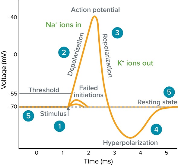
An action potential is a rapid rise and subsequent fall in voltage or membrane potential across a cellular membrane with a characteristic pattern. Sufficient current is required to initiate a voltage response in a cell membrane; if the current is insufficient to depolarize the membrane to the threshold level, an action potential will not fire. Examples of cells that signal via action potentials are neurons and muscle cells. 
Action Potential Graph
  1. Stimulus starts the rapid change in voltage or action potential. In patch-clamp mode, sufficient current must be administered to the cell in order to raise the voltage above the threshold voltage to start membrane depolarization.
  2. Depolarization is caused by a rapid rise in membrane potential opening of sodium channels in the cellular membrane, resulting in a large influx of sodium ions.
  3. Membrane Repolarization results from rapid sodium channel inactivation as well as a large efflux of potassium ions resulting from activated potassium channels.
  4. Hyperpolarization is a lowered membrane potential caused by the efflux of potassium ions and closing of the potassium channels.
  5. Resting state is when membrane potential returns to the resting voltage that occurred before the stimulus occurred.

MRI Machine

Magnetic resonance imaging (MRI), also known as nuclear magnetic resonance imaging, is a scanning technique for creating detailed images of the human body.
The scan uses a strong magnetic field and radio waves to generate images of parts of the body that can't be seen as well with X-rays, CT scans or ultrasound. For example, it can help doctors to see inside joints, cartilage, ligaments, muscles and tendons, which makes it helpful for detecting various sports injuries.
MRI is also used to examine internal body structures and diagnose a variety of disorders, such as strokes, tumours, aneurysms, spinal cordinjuries, multiple sclerosis and eye or inner ear problems, t is also widely used in research to measure brain structure and function, among other things. 
"What makes MRI so powerful is, you have really exquisite soft tissue, and anatomic, detail," The biggest benefit of MRI compared with other imaging techniques (such as CT scans and x-rays) is, there's no risk of being exposed to radiation, Filippi told Live Science.
During an MRI, a person will be asked to lie on a movable table that will slide into a doughnut-shaped opening of the machine to scan a specific portion of your body. The machine itself will generate a strong magnetic field around the person and radio waves will be directed at the body.
A person will not feel the magnetic field or radio waves, so the procedure itself is painless. However, there may be a lot of loud thumping or tapping noises during the scan (it may sound like a sledgehammer!), so people are often given headphones to listen to music or earplugs to help block the sound. A technician may also give instructions to you during the test. 
Some people may be given a contrast solution by intravenous, a liquid dye that can highlight specific problems that might not show up otherwise on the scan. 
Young children as well as people who feel claustrophobic in enclosed places may be given sedating medication to help them relax or fall asleep during the scan because it is important to stay as still as possible to get clear images. Movement can blur the images. 
Some hospitals might have an open MRI machine that is open on the sides rather than the tunnel-like tube found in a traditional machine. This may be a helpful alternative for people who feel afraid of confined spaces. 
The scan itself may take 30 to 60 minutes, on average, according to the American Academy of Family Physicians.
A radiologist will look at the images and send a report to your doctor with your test results. 
The human body is mostly water. Water molecules (H2O) contain hydrogen nuclei (protons), which become aligned in a magnetic field. An MRI scanner applies a very strong magnetic field (about 0.2 to 3 teslas, or roughly a thousand times the strength of a typical fridge magnet), which aligns the proton "spins."
Image result for MRI MACHINE BLOCK DIAGRAM
The scanner also produces a radio frequency current that creates a varying magnetic field. The protons absorb the energy from the magnetic field and flip their spins. When the field is turned off, the protons gradually return to their normal spin, a process called precession. The return process produces a radio signal that can be measured by receivers in the scanner and made into an image.
An MRI scan reveals the gross anatomical structure of the human brain.
An MRI scan reveals the gross anatomical structure of the human brain
Protons in different body tissues return to their normal spins at different rates, so the scanner can distinguish among various types of tissue. The scanner settings can be adjusted to produce contrasts between different body tissues. Additional magnetic fields are used to produce 3-dimensional images that may be viewed from different angles. There are many forms of MRI, but diffusion MRI and functional MRI (fMRI) are two of the most common.
This form of MRI measures how water molecules diffuse through body tissues. Certain disease processes — such as a stroke or tumor — can restrict this diffusion, so this method is often used to diagnose them.
Functional MRI
In addition to structural imaging, MRI can also be used to visualize functional activity in the brain. Functional MRI, or fMRI, measures changes in blood flow to different parts of the brain. 
It is used to observe brain structures and to determine which parts of the brain are handling critical functions. Functional MRI may also be used to evaluate damage from a head injury or Alzheimer's disease. fMRI has been especially useful in neuroscience — "It has really revolutionized how we study the brain,"
Unlike other imaging forms like X-rays or CT scans, MRI doesn't use ionizing radiation. MRI is increasingly being used to image fetuses during pregnancy, and no adverse effects on the fetus have been demonstrated.
Still, the procedure can have risks, and medical societies don't recommend using MRI as the first stage of diagnosis.
Because MRI uses strong magnets, any kind of metal implant, such as a pacemaker, artificial joints, artificial heart valves, cochlear implants or metal plates, screws or rods, pose a hazard. The implant can move or heat up in the magnetic field. 
Several patients with pacemakers who underwent MRI scans have died, patients should always be asked about any implants before getting scanned. Many implants today are "MR-safe,"
The constant flipping of magnetic fields can produce loud clicking or beeping noises, so ear protection is necessary during the scan.